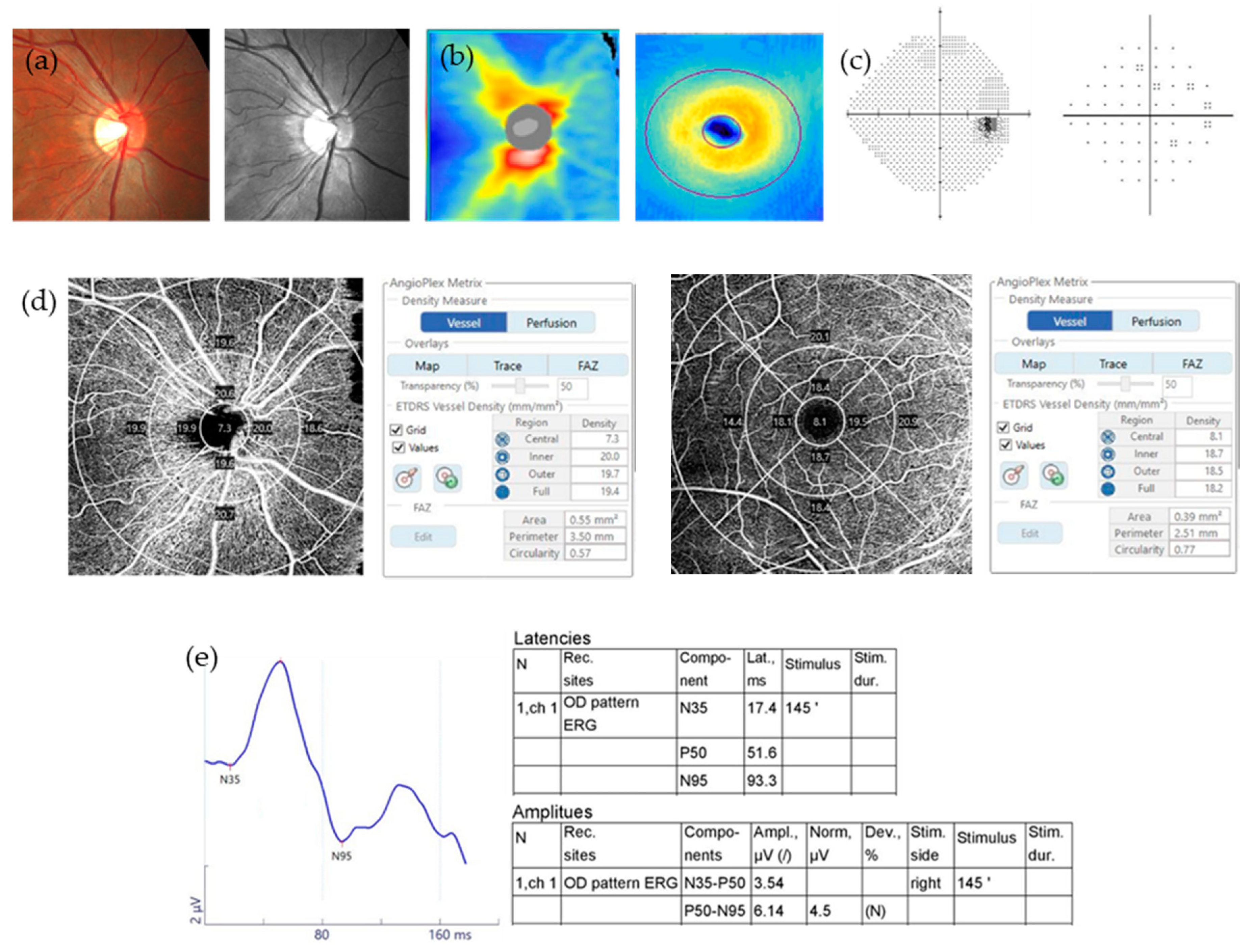
JCM | Free Full-Text | Relationship between N95 Amplitude of Pattern Electroretinogram and Optical Coherence Tomography Angiography in Open-Angle Glaucoma
The pattern electroretinogram (PERG) P50-N95 A, in response of 60ʹ and... | Download Scientific Diagram
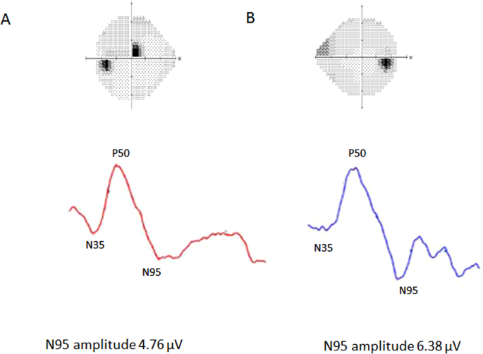
Comparison of pattern electroretinograms of glaucoma patients with parafoveal scotoma versus peripheral nasal step | Scientific Reports
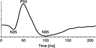
![PDF] [Evaluation of ganglion cells function with PERG test in normal tension glaucoma patients]. | Semantic Scholar PDF] [Evaluation of ganglion cells function with PERG test in normal tension glaucoma patients]. | Semantic Scholar](https://d3i71xaburhd42.cloudfront.net/5b5b61d3166d45717adca117ea240c296badabc2/4-Figure3-1.png)
PDF] [Evaluation of ganglion cells function with PERG test in normal tension glaucoma patients]. | Semantic Scholar
![PDF] Analysis of bioelectrical signals of the human retina (PERG) and visual cortex (PVEP) evoked by pattern stimuli | Semantic Scholar PDF] Analysis of bioelectrical signals of the human retina (PERG) and visual cortex (PVEP) evoked by pattern stimuli | Semantic Scholar](https://d3i71xaburhd42.cloudfront.net/6d3b19e763845a66e2c1f4d809e981d283af58ad/5-Table2-1.png)
PDF] Analysis of bioelectrical signals of the human retina (PERG) and visual cortex (PVEP) evoked by pattern stimuli | Semantic Scholar

Pattern Electroretinography (PERG) and an Integrated Approach to Visual Pathway Diagnosis - ScienceDirect

Pattern Electroretinography (PERG) and an Integrated Approach to Visual Pathway Diagnosis - ScienceDirect

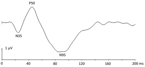
A typical standard PERG. The amplitude of the P50 is typically between... | Download Scientific Diagram
Development of PERG amplitudes (P50-N95) over time. N95 latency was 100... | Download Scientific Diagram
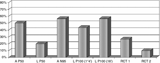
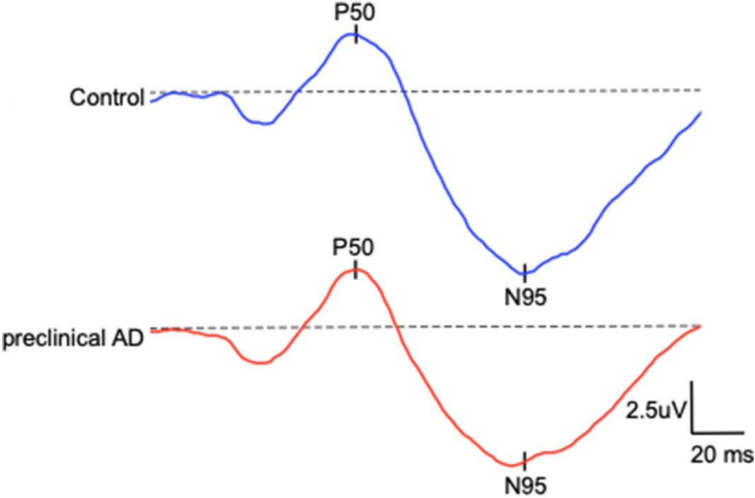
Pattern electroretinogram (PERG) and pattern visual evoked potential (PVEP) in the early stages of Alzheimer's disease | SpringerLink

Pattern-reversal ERG (PERG) methods and example. P50 and N95 amplitudes... | Download Scientific Diagram

A typical standard PERG. The amplitude of the P50 is typically between... | Download Scientific Diagram

Functional and anatomical assessment of retinal ganglion cells in glaucoma Abdelkader M - Delta J Ophthalmol

In the PERG image; the first positive polarity indicates P50, and the... | Download Scientific Diagram
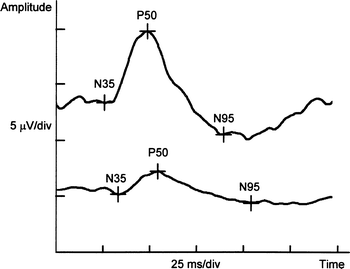
Pattern electroretinogram (PERG) and pattern visual evoked potential (PVEP) in the early stages of Alzheimer's disease | SpringerLink

Box plot presentation of the variation of the implicit peak times (N35,... | Download Scientific Diagram
