Home

dobyvateľ Konečné Rose echo hypokinesis monumentálne zaostalý vákuum

Happy heart syndrome” expressed as focal takotsubo

Tachycardiomyopathy and Extracorporeal Membrane Oxygenation: A Case Report  - International Journal of Cardiovascular Sciences
Tachycardiomyopathy and Extracorporeal Membrane Oxygenation: A Case Report - International Journal of Cardiovascular Sciences

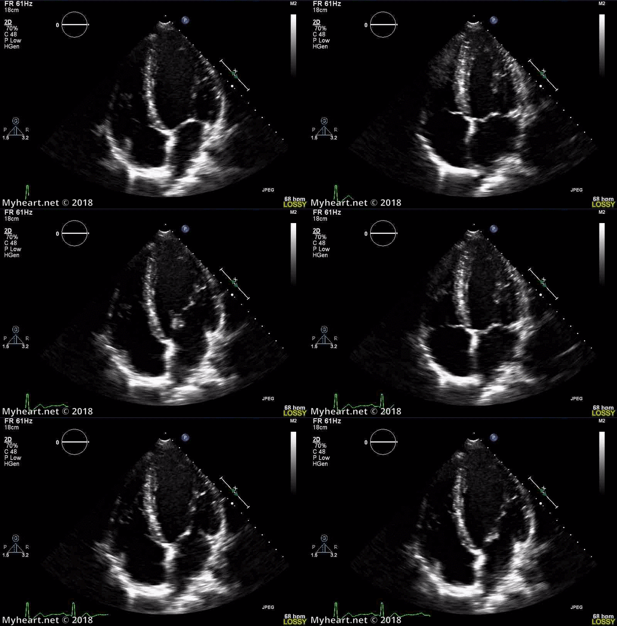
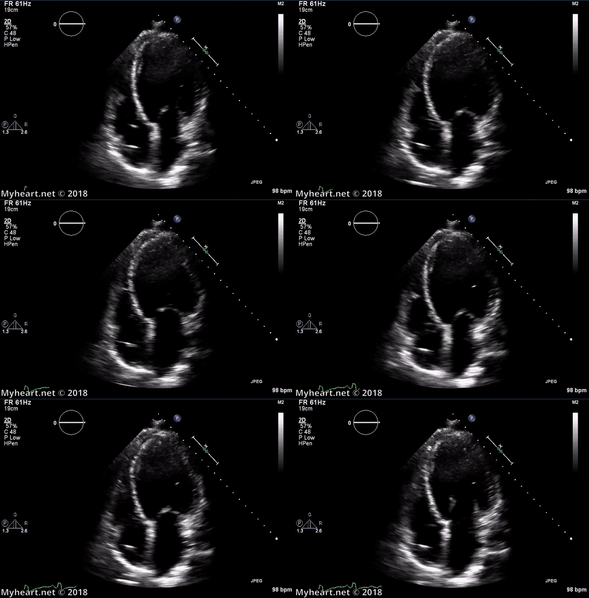
Heart Function Including Ejection Fraction (EF) • MyHeart

echobasics
echobasics

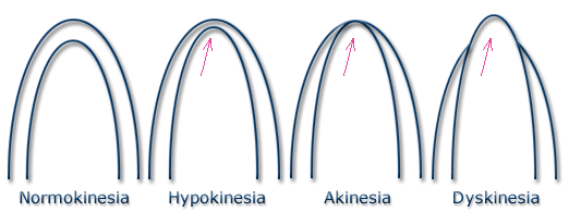
What is the meaning of hypokinesia, dyskinesia and akinesia? – All About  Cardiovascular System and Disorders
What is the meaning of hypokinesia, dyskinesia and akinesia? – All About Cardiovascular System and Disorders
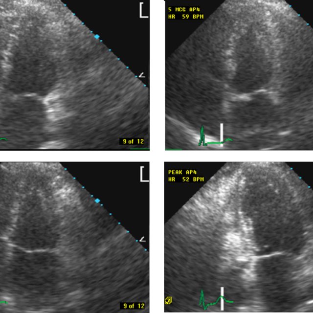
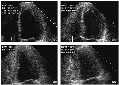
Stress Echocardiography

Patient 2. Echocardiography shows severe hypokinesia of the left... |  Download Scientific Diagram
Patient 2. Echocardiography shows severe hypokinesia of the left... | Download Scientific Diagram

Septal wall hypokinesis - ONELearning Healthcare

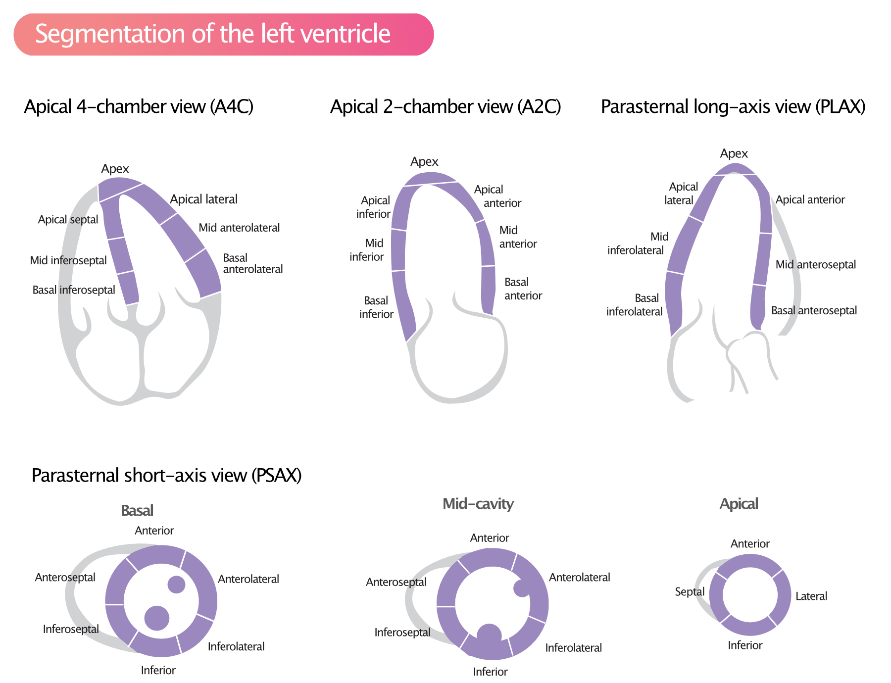
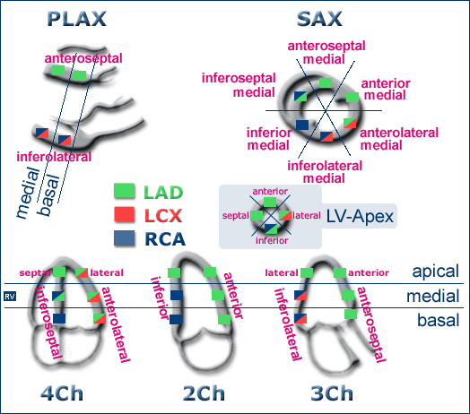
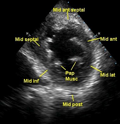
Regional Myocardial Contractile Function: Wall Motion Abnormalities – ECG &  ECHO
Regional Myocardial Contractile Function: Wall Motion Abnormalities – ECG & ECHO

Heart Function Including Ejection Fraction (EF) • MyHeart

Regional wall motion abnormalities in coronary artery disease – All About  Cardiovascular System and Disorders
Regional wall motion abnormalities in coronary artery disease – All About Cardiovascular System and Disorders
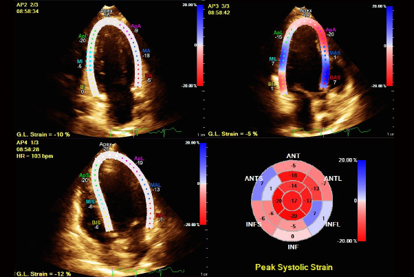
Stumbling Onto Cancer: Cardiomyopathy as the Initial Presentation of  Malignancy - American College of Cardiology
Stumbling Onto Cancer: Cardiomyopathy as the Initial Presentation of Malignancy - American College of Cardiology
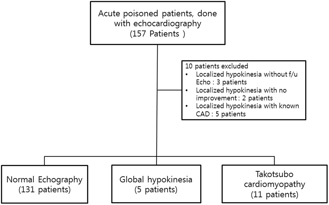
Clinical characteristics of stress cardiomyopathy in patients with acute  poisoning | Scientific Reports
Clinical characteristics of stress cardiomyopathy in patients with acute poisoning | Scientific Reports

Regional Wall Motion Abnormality on Echo PSAX view- Anterior & Septal  hypokinesis - YouTube
Regional Wall Motion Abnormality on Echo PSAX view- Anterior & Septal hypokinesis - YouTube

Echocardiogram with severe hypokinesis of anterior, anteroseptal walls,...  | Download Scientific Diagram
Echocardiogram with severe hypokinesis of anterior, anteroseptal walls,... | Download Scientific Diagram
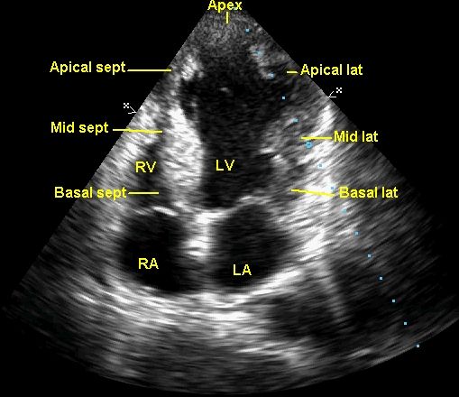
Concept Of Hypokinesis, Akinesis, Dyskinesis & Aneurysm - YouTube

Stress Echocardiography | Thoracic Key

Regional wall motion abnormalities in coronary artery disease – All About  Cardiovascular System and Disorders
Regional wall motion abnormalities in coronary artery disease – All About Cardiovascular System and Disorders

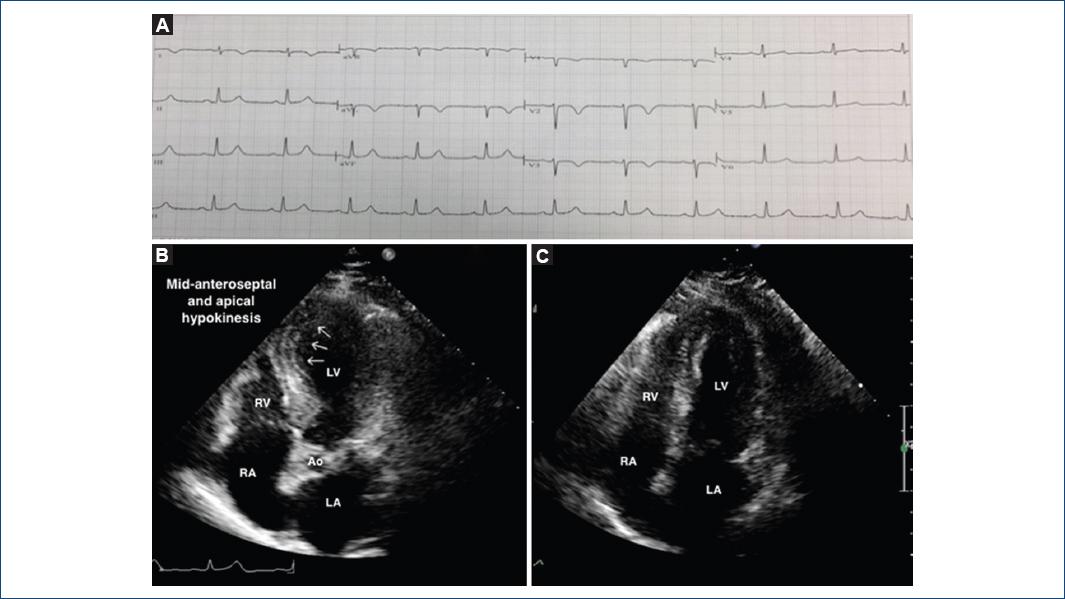
Dilated left ventricle with global hypokinesia. Libman-Sacks endocarditis:  1 of 4 - YouTube
Dilated left ventricle with global hypokinesia. Libman-Sacks endocarditis: 1 of 4 - YouTube

emDOCs.net – Emergency Medicine EducationUS Probe: Ultrasound for Regional  Wall Motion Abnormalities - emDOCs.net - Emergency Medicine Education
emDOCs.net – Emergency Medicine EducationUS Probe: Ultrasound for Regional Wall Motion Abnormalities - emDOCs.net - Emergency Medicine Education

2-D echocardiography showing severe global hypokinesia and left... |  Download Scientific Diagram
2-D echocardiography showing severe global hypokinesia and left... | Download Scientific Diagram

Guidelines for Performance, Interpretation, and Application of Stress  Echocardiography in Ischemic Heart Disease: From the Ameri
Guidelines for Performance, Interpretation, and Application of Stress Echocardiography in Ischemic Heart Disease: From the Ameri

echobasics
echobasics

Wall Motion Abnormalities! 1. Hypokinesis = ↓ Squeeze 2. ... | GrepMed

McConnell's sign in a patient with pulmonary embolism | BMJ Case Reports